Facilitates and Labs
We have a lot of facilities and labs here at Princeton. Check out this list to see how you can access them:At Princeton, we have a lot of facilities and labs. If you’re interested in studying the human brain, our Neuroscience Institute is the place for you. Our organization has a specialized facility called the Centre for Molecular and Cellular Analysis (CMCA). This facility conducts biomedical research using advanced techniques like genomics and proteomics. If your interests lie more with chemistry or biology than neuroscience, check out our Chemistry Department’s Analytical Laboratory or Biochemistry Department’s Protein Expression Laboratory!

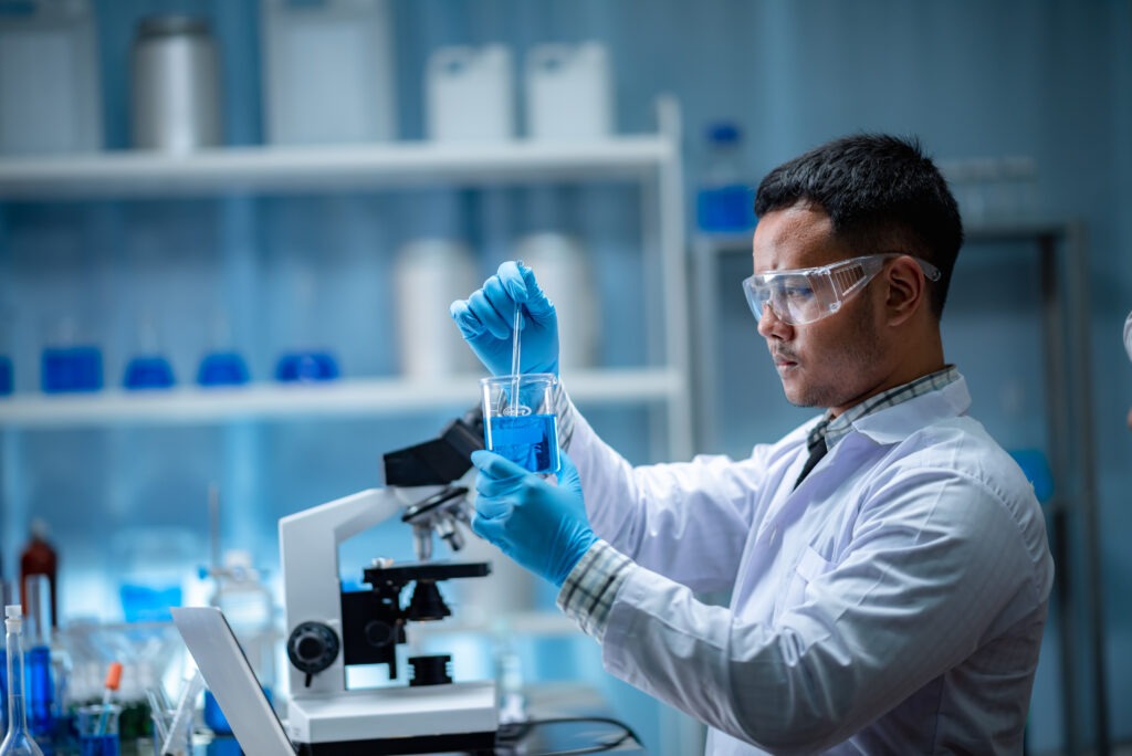
The Centre for Molecular and Cellular Analysis (CMCA)
The Centre for Molecular and Cellular Analysis (CMCA) is a research facility at Princeton University. It houses several essential facilities, including the following:
- Protein Expression and Purification Core Facility
- NMR Spectroscopy Core Facility
- Fluorescence Microscopy and Imaging Centre (FMIC)
This centre also hosts many other resources that you may find helpful, including an Applied Biotechnology Lab and a Mass Spectrometry Facility.
The Imaging and Analysis Centre (IAC)
The Imaging and Analysis Centre (IAC) is a shared facility for the Princeton Neuroscience Institute and the Department of Molecular Biology. The facility has advanced confocal microscopes, fluorescence microscopes, and fluorescence stereomicroscopes. Expert neurobiologists, biochemists, and molecular biologists operate these instruments.
The IAC provides access to advanced imaging techniques and expertise in their use–all at no cost to students or faculty!

The Neuroscience Institute
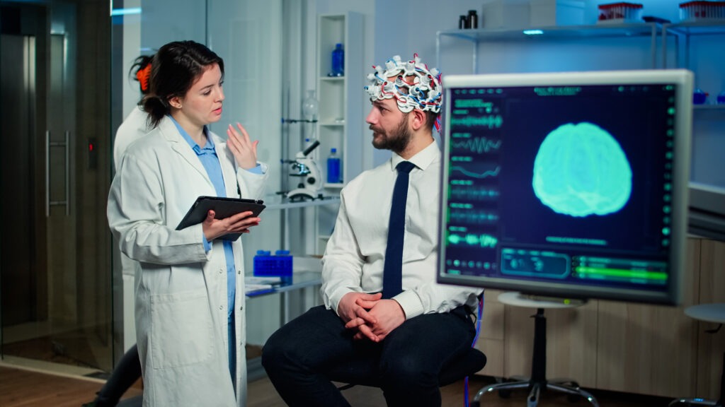
The Neuroscience Institute was established in 1991 to facilitate collaboration among Princeton’s neuroscience researchers. It is a consortium of neuroscience researchers from the University and the Medical Centre who work together on projects ranging from studies of brain function, behaviour and development to clinical research involving patients with neurological disorders.
The Neuroscience Institute provides shared resources for its members’ research efforts: facilities such as the Brain Imaging Centre (BIC), which houses state-of-the-art MRI scanners; an animal facility where mice can be genetically engineered or trained; an electron microscopy suite; a tissue culture room where cells can be grown outside their natural environment; and several core facilities that provide support services such as genotyping or cell culture preparation.
The S.U.N.Y.-ESF Genomics Core Facility (GCF)
The S.U.N.Y.-ESF Genomics Core Facility (GCF) is a state-of-the-art genomic research and education facility. It has equipment for DNA sequencing, genotyping, and next-generation sequencing. In addition, they also have equipment for isolating and purifying DNA and other tools used in the field of genetics research, such as PCR machines or gel electrophoresis systems. Their staff consists of experts who can teach you how to use their equipment or do the work for you if needed!
The GCF website has much more information about their services, so check it out if interested!

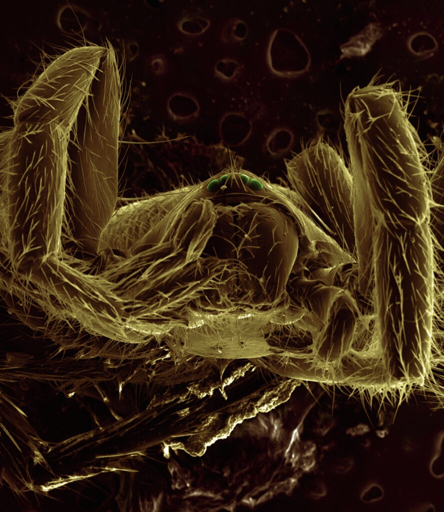
The Confocal Microscopy Facility (CMF)
The Confocal Microscopy Facility (CMF) is a state-of-the-art facility for confocal microscopy. The CMF is equipped with the latest confocal microscope equipment, including Zeiss LSM 780/880 and Zeiss Axiovert 200M inverted microscopes, both equipped with gradient index (GRIN) lenses and lasers for laser scanning confocal microscopy (LSCM).
The Confocal Microscopy Facility also has two custom-built LSCM systems in which the imaging components are mounted on an inverted microscope platform. These systems allow us to take advantage of all aspects of LSCM technology, including the simultaneous acquisition of multiple planes through different combinations of pinholes or beamsplitters; widefield imaging using low numerical aperture objectives; use of long working distances without compromising resolution; high-speed imaging via fast shutters/shutters etc.